Picture a city where all the traffic flows through a tangle of expressways, a place where off-ramps to the neighborhoods and side streets don’t exist.
Imagine the pressure this puts on the main thoroughfares.
Not an ideal situation.
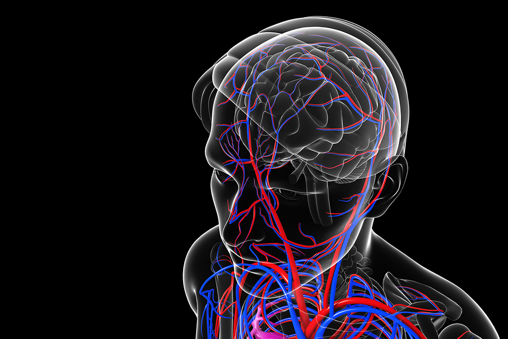
That’s kind of what it’s like in the brain of a person with a cerebral arteriovenous malformation, or AVM, an abnormal connection of major veins and arteries joined in a way that bypasses the capillaries. There’s no step-down system for the blood vessels in that part of the brain.
“Your normal plumbing is (big) arteries to smaller arteries to capillaries to tissue, to really small veins to bigger veins to major veins,” explained Justin Singer, MD, Spectrum Health Medical Group’s director of vascular neurosurgery, who specializes in minimally invasive neuro-endovascular surgery.
With an AVM, however, the blood isn’t allowed to spread out and disperse through this branching system of veins and arteries. Instead, it’s funneled into an abnormal tangle of vessels with no outlet to the brain tissue.
“And that causes high pressure and increases the risk of these bad arteries hemorrhaging,” Dr. Singer said.
Specialized treatment
AVMs are rare, affecting less than 1 percent of the population, according to the American Stroke Association. Because they require specialized expertise, the malformations are typically treated at major stroke centers.
The Spectrum Health Stroke Center sees about 20 AVM cases a year. Over a two-month period in early 2017, Dr. Singer treated five AVM patients, including two children.
The neurovascular team treats children in collaboration with pediatric neurology specialists from Spectrum Health Helen DeVos Children’s Hospital. In March, Health Beat published the AVM survival story of Brianna Laux, age 12.
AVMs are congenital and typically start out silent—without symptoms, Dr. Singer said. Patients can live for decades without knowing they have one.
Then, often in their teens, 20s or 30s, these patients might have a bad headache or a small seizure and go to the hospital to have it checked out.
That’s when they learn that they have an AVM—and that it has ruptured. Most AVMs have a 2 to 5 percent risk of hemorrhaging per year. There’s also the risk that an aneurysm can develop in the middle of an AVM.
If the hemorrhage is large, it may need immediate intervention. If the bleed is smaller, doctors may have a day or two to explore their options.
AVMs are by nature complex and treatment varies based on their location in the brain and their level of risk. Depending on the case, the neurovascular team may recommend any of the following approaches to treatment:
- Observation—If the AVM is in a tricky part of the brain and the risk of doing surgery or other intervention outweighs the risk of simply monitoring the patient, doctors take a nonaggressive approach.
- Stereotactic radiosurgery, or SRS—This treatment aims an intense dose of radiation at a specific part of the AVM to obliterate it or shrink it to a size that will be safer to operate on later.
- Surgery to remove the AVM—If the abnormal vessels are in a place that’s easy to reach, surgery may be the best option. The neurosurgeon removes part of the patient’s skull, then carefully removes the AVM, seals off the vessels and replaces the skull piece. High-tech fluorescent microscopes in the operating room allow surgeons to inject dye and “light up” the vessels they need to target.
- Endovascular embolization—This advanced procedure uses a tiny catheter, inserted through an artery in the groin, to deliver a glue-like substance called Onyx into the AVM to block it and stop the bleeding. Real-time brain scans let the surgeons track and direct the flow of the glue. Sometimes the embolization serves as an alternative to surgery, to prevent a stroke. But most of the time, embolization is used as a prelude to surgery. “If we can do it well, we make the surgical removal of the AVM much easier,” Dr. Singer said.
Innovations
Though endovascular embolization isn’t a new procedure, Dr. Singer and his Spectrum Health colleagues are always working to improve their methods and outcomes.
“We’ve been very successful recently in optimizing our techniques to get (the Onyx) to penetrate where we want it to go,” he said. One new trick uses a special balloon to help control the path of the glue, which flows a bit like lava.
Dr. Singer is part of a multidisciplinary team at Spectrum Health that includes interventional radiologists, neurosurgeons, endovascular neurosurgeons, neurologists, vascular neurologists and other neuro critical care doctors, along with a dedicated neuro intensive care unit.
“We have multiple knowledge sources to put our heads together to decide what’s best for every patient,” he said. “It’s very much a team effort.”
Dr. Singer is excited to be part of what he calls a “revolution and renaissance” in the neurovascular field.
“We’re making great advances across all of what we treat in neurovascular, whether it’s aneurysms or AVMs,” he said.
The new treatments coming down the line—new radiation therapies, surgical techniques, medications, glues and other catheter-based technologies—“will help make some of these really bad pathologies more curable in less invasive ways,” he said.
The end game is to minimize patient risk in a super-risky field.
 /a>
/a>
 /a>
/a>
 /a>
/a>