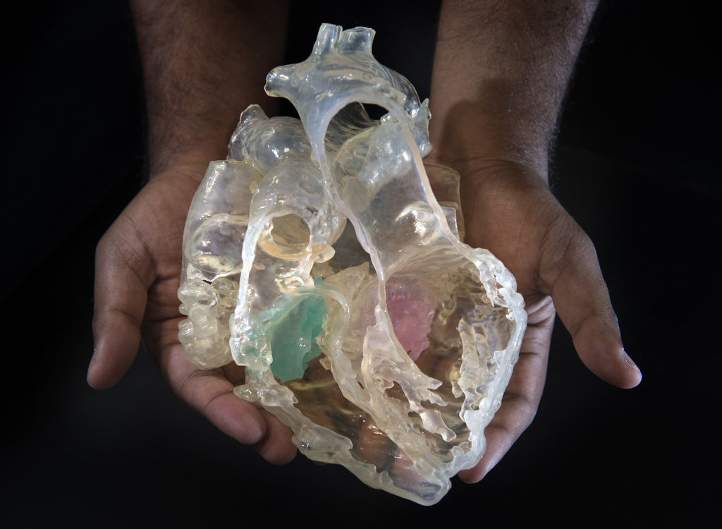
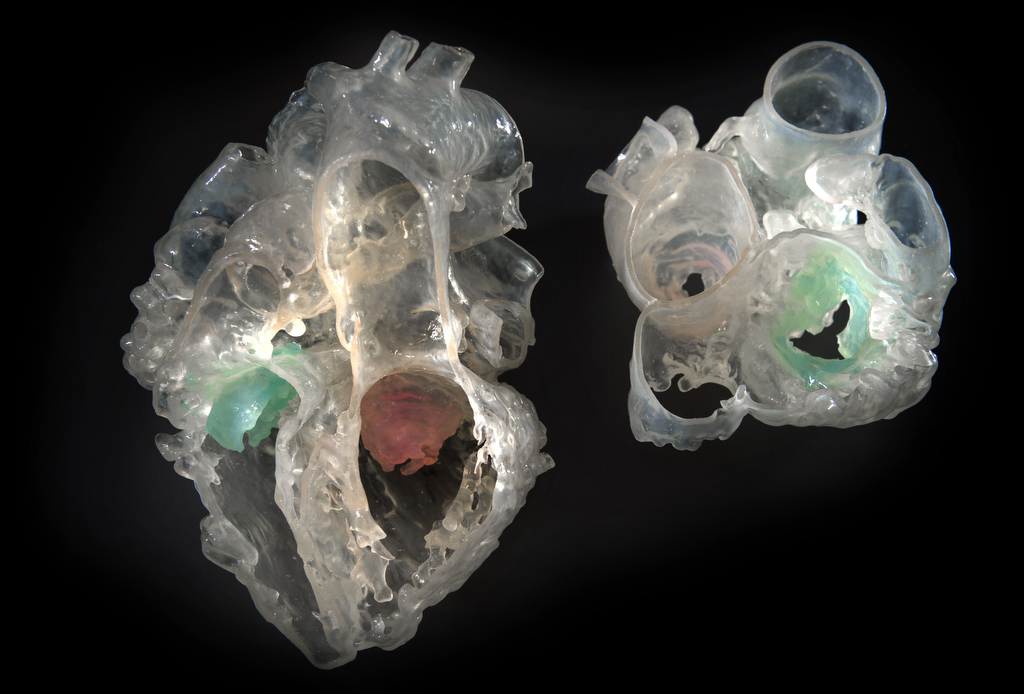
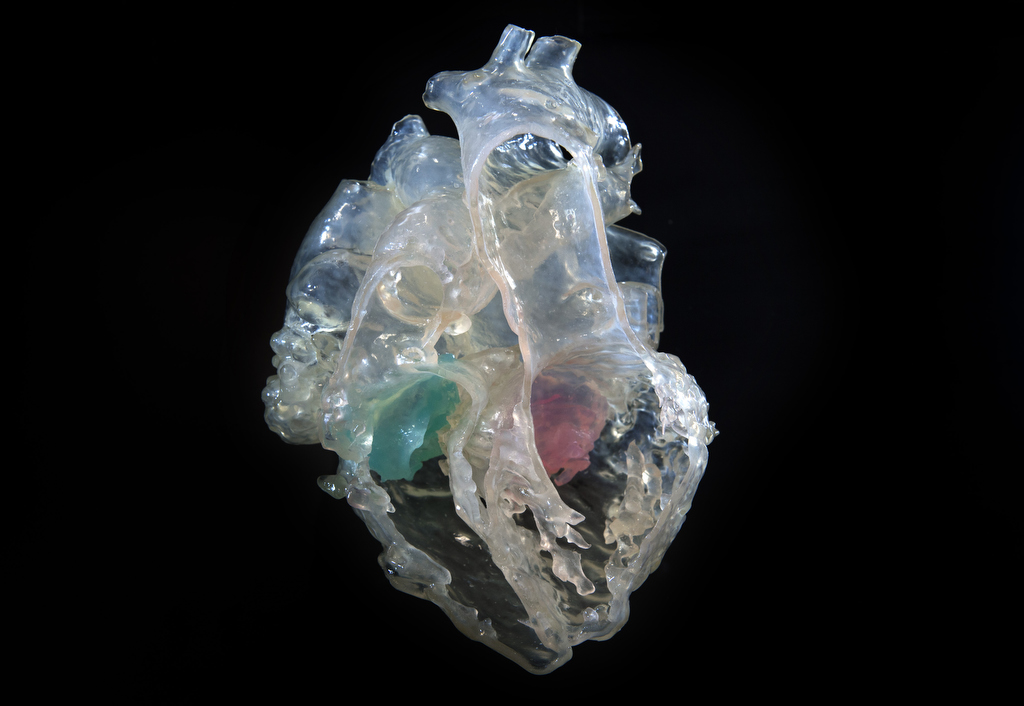
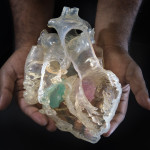
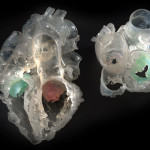
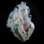
Congenital heart experts from Spectrum Health Helen DeVos Children’s Hospital recently integrated two common imaging techniques for the first time to produce a more complete and accurate three-dimensional printed model of a patient’s heart.
This advancement provides heart surgeons and cardiologists with a tool to better visualize, diagnose and treat diseases, for which they once had to rely on two-dimensional imaging or invasive exploratory surgeries.
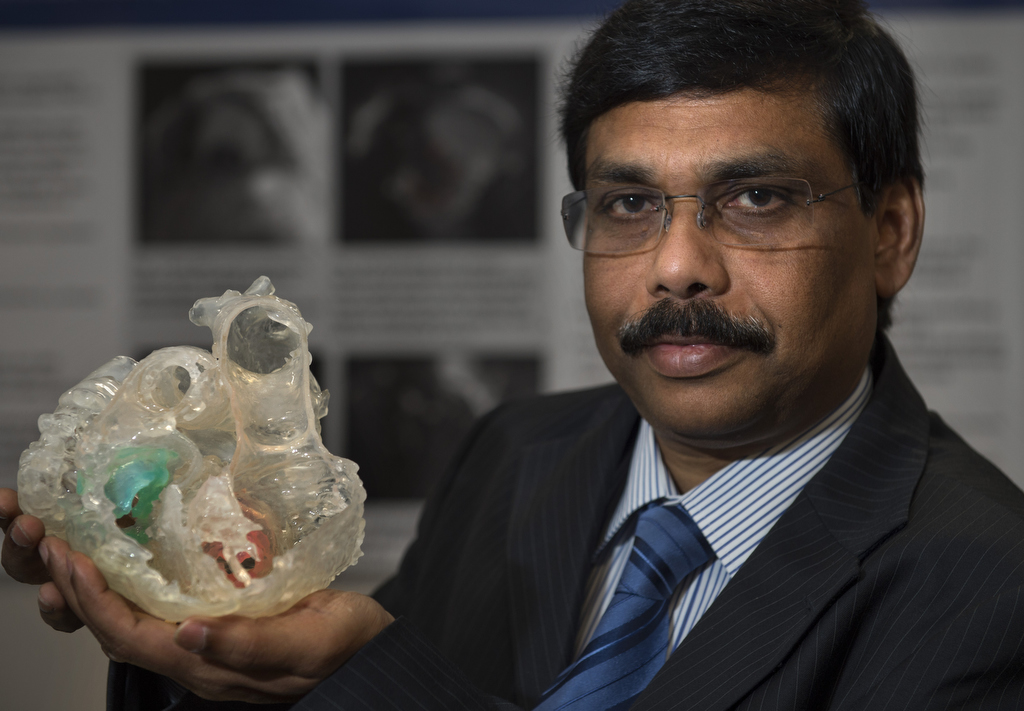
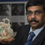
“This is a huge leap for individualized medicine in cardiology and congenital heart disease,” said Joseph Vettukattil, MD, a pediatric cardiologist and co-director of the Helen DeVos Children’s Hospital Congenital Heart Center. Dr. Vettukattil is senior author of a study on the topic, which he recently presented in Frankfurt, Germany.
“Before you open the patient’s chest, and the heart, you already have a clear spatial understanding of the structures in the heart and how you are going to address the defect,” Dr. Vettukattil said.
According to the Centers for Disease Control and Prevention, more than 600,000 people die of heart disease in the United States every year–that’s 1 in every 4 deaths and more than all types of cancer combined.
Three-dimensional printing of patients’ hearts has become more common in recent years as part of an emerging, experimental field devoted to visualizing individual cardiac structures and characteristics.
But this is the first time the integration of computed tomography, more commonly known as a CT scan, and 3-D transesophageal echocardiography has successfully been used for printing a hybrid 3-D model of a patient’s heart.
The study also opens the way for these techniques to be used in combination with a third tool–magnetic resonance imaging, commonly known as an MRI.
“Hybrid 3-D printing integrates the best aspects of two or more imaging modalities, which can potentially enhance diagnosis, as well as interventional and surgical planning,” said Jordan Gosnell, Helen DeVos Children’s Hospital cardiac sonographer, and lead author of the study. “Previous methods of 3-D printing utilize only one imaging modality, which may not be as accurate.”
According to Dr. Vettukattil and Gosnell, each imaging tool has different strengths, which can improve and enhance 3-D printing:
- CT enhances visualization of the outside anatomy of the heart
- MRI is superior to other imaging techniques for measuring the interior of the heart, including the right and left ventricles or main chambers of the heart, as well as the heart’s muscular tissue
- 3-D transesophageal echocardiography provides the best visualization of valve anatomy
CT and MRI are established imaging tools for producing 3-D printable models. Dr. Vettukattil and his team recently reported the third scan to be a feasible imaging technique to generate 3-D printing in congenital heart disease.
Congenital heart defects form prior to birth.
They can take many forms, occurring in the heart’s chambers, valves or blood vessels. Some may be life-threatening at birth, while others may develop slowly over a lifetime.
Common problems include holes in the heart, narrowing of the pulmonary and aortic valves, narrowing of the aorta, the heart’s main artery, and abnormal connections of blood vessels with the heart. This model was produced from a patient with complex congenital heart disease.
The team is currently working on another patient’s 3-D heart model. The printing process takes place in Belgium at the headquarters of the team’s study partner, Materialise NV, a leading provider of 3-D printing software and services, which prints the model using its HeartPrint Flex technology.
In addition to diagnosis and treatment, the models also serve another important function–that of education.
“The 3-D heart may also be used to teach patients and their family members about their congenital heart defect and the plan for repair,” said Bennett Samuel, MHA, BSN, another of the study’s authors and clinical research nurse at the Helen DeVos Children’s Hospital Congenital Heart Center. “Currently a two-dimensional representation, such as a drawing on a piece of paper or whiteboard, is used to explain the procedure to the patient.”
“Finally, the 3-D printed models may be beneficial to teach medical students, residents, nurses and other medical professionals about specific congenital heart defects,” he added.
 /a>
/a>
 /a>
/a>
 /a>
/a>
Really like these articles about congenital heart disease, as I was born with Tettrology of Fallott. At the age of 18 I had open heart surgery and was given a 50/50 chance of surviving the surgery. I am 71 years old and living with the help of my 13th pacemaker for 3rd degree heart block. What a long way we have come since my birth!
Wow, Carol! What an awesome story and so glad you’re doing so well. Thanks for being a Health Beat reader! If you haven’t already, definitely bookmark our website on your phone and computer – spectrumhealthbeat.org – and then come back to us often as we update our website with new stories you may enjoy every single day. 🙂